2013


C. Jones, M. Seyedhosseini, M. Ellisman, T. Tasdizen.
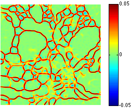
“Neuron Segmentation in Electron Microscopy Images Using Partial Differential Equations,” In Proceedings of 2013 IEEE 10th International Symposium on Biomedical Imaging (ISBI), pp. 1457--1460. April, 2013.
DOI: 10.1109/ISBI.2013.6556809
K.B. Jones, M. Datar, S. Ravichandran, H. Jin, E. Jurrus, R.T. Whitaker, M.R. Capecchi.
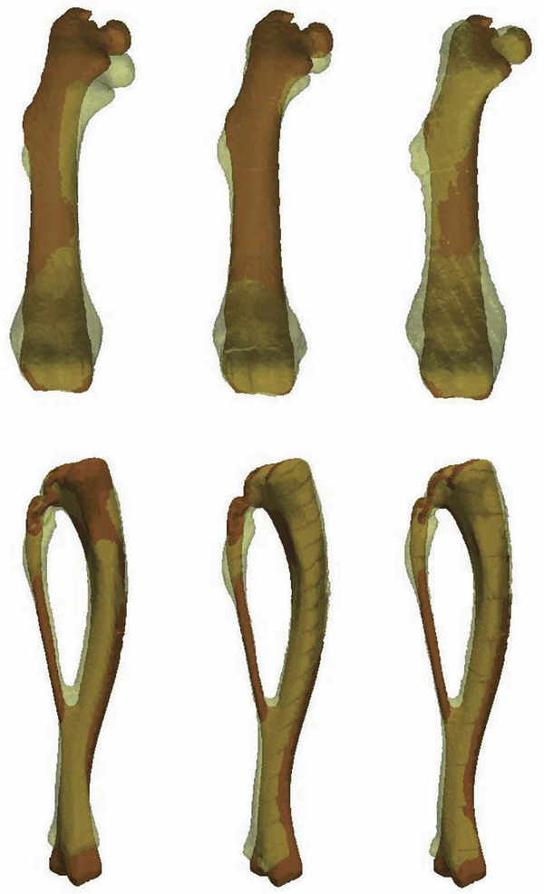
“Toward an Understanding of the Short Bone Phenotype Associated with Multiple Osteochondromas,” In Journal of Orthopaedic Research, Vol. 31, No. 4, pp. 651--657. 2013.
DOI: 10.1002/jor.22280
PubMed ID: 23192691
PubMed Central ID: PMC3683979
2010
J.R. Anderson, B.C. Grimm, S. Mohammed, B.W. Jones, T. Tasdizen, J. Spaltenstein, P. Koshevoy, R.T. Whitaker, R.E. Marc.
“The Viking Viewer: Scalable Multiuser Annotation and Summarization of Large Volume Datasets,” In Journal of Microscopy, Vol. 241, No. 1, pp. 13--28. 2010.
DOI: 10.1111/j.1365-2818.2010.03402.x
2008
S.X. Vasquez, M.S. Hansen, A.N. Bahadur, M.F. Hockin, G.L. Kindlmann, L. Nevell, I.Q. Wu, D.J. Grunwald, D.M. Weinstein, G.M. Jones, C.R. Johnson, J.L. Vandeberg, M.R. Capecchi, C. Keller.
“Optimization of Volumetric Computed Tomography for Skeletal Analysis of Model Genetic Organisms,” In The Anatomical Record: Advances in Integrative Anatomy and Evolutionary Biology, Vol. 291, pp. 475--487. 2008.
PubMed ID: 18286615
2006
S. Browd, L.J. Healy, G. Dobie, J.T. Johnson III, G.M. Jones, L.F. Rodriguez, D.L. Brockmeyer.
“Morphometric and Qualitative Analysis of Congenital Occipitocervical Instability in Children: Implications for Down Syndrome Patients,” In Journal of Neurosurgery: Pediatrics, Vol. 105, No. 1 , Journal of Neurosurgery Publishing Group, pp. 50--54. July, 2006.
DOI: 10.3171/ped.2006.105.1.50
J.T. Johnson III, M.S. Hansen, I. Wu, L.J. Healy, C.R. Johnson, G.M. Jones, M.R. Capecchi, C. Keller.
“Virtual Histology of Transgenic Mouse Embryos for High-Throughput Phenotyping,” In PLoS Genetics, Vol. 2, No. 1, pp. 471--477. April, 2006.
J.T. Johnson III, M.S. Hansen, I.Q Wu, L.J. Healy, C.R. Johnson, G.M. Jones, M.R. Capecchi, C.Keller.
“Virtual Histology of Transgenic Mouse Embryos for High-Throughput Phenotyping,” In Proceedings of the Teratology 46th Annual Meeting, Tucson, AZ, 2006.
2005
P. Hawkes, G. Kindlmann, D. Weinstein, G.M. Jones, C. Keller.
“Practical Vessel Imaging by Computed Tomography in Live Transgenic Mouse Models for Human Tumors,” In Proceedings of 2005 SMI Conference, 4th Annual Meeting of the Society for Molecular Imaging, Cologne, Germany, 2005.
G.L. Kindlmann, D.M. Weinstein, G.M. Jones, C.R. Johnson, M.R. Capecchi, C. Keller.
“Practical Vessel Imaging by Computed Tomography in Live Transgenic Mouse Models for Human Tumors,” In Journal of Molecular Imaging, Vol. 4, No. 4, pp. 417--424. 2005.
2004
J. Cates, R.T. Whitaker, G.M. Jones.
“Case Study: An Evaluation of User-Assisted Hierarchical Watershed Segmentation,” No. UUCS-04-006, University of Utah School of Computing, 2004.