
Image Analysis
SCI's imaging work addresses fundamental questions in 2D and 3D image processing, including filtering, segmentation, surface reconstruction, and shape analysis. In low-level image processing, this effort has produce new nonparametric methods for modeling image statistics, which have resulted in better algorithms for denoising and reconstruction. Work with particle systems has led to new methods for visualizing and analyzing 3D surfaces. Our work in image processing also includes applications of advanced computing to 3D images, which has resulted in new parallel algorithms and real-time implementations on graphics processing units (GPUs). Application areas include medical image analysis, biological image processing, defense, environmental monitoring, and oil and gas.
Ross Whitaker
Segmentation

Chris Johnson
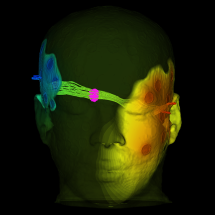
Diffusion Tensor Analysis
Funded Research Projects:
Publications in Image Analysis:
  A fast iterative method for solving the Eikonal equation on triangulated surfaces Z. Fu, W.-K. Jeong, Y. Pan, R.M. Kirby, R.T. Whitaker. In SIAM Journal of Scientific Computing, Vol. 33, No. 5, pp. 2468--2488. 2011. DOI: 10.1137/100788951 PubMed Central ID: PMC3360588 This paper presents an efficient, fine-grained parallel algorithm for solving the Eikonal equation on triangular meshes. The Eikonal equation, and the broader class of Hamilton–Jacobi equations to which it belongs, have a wide range of applications from geometric optics and seismology to biological modeling and analysis of geometry and images. The ability to solve such equations accurately and efficiently provides new capabilities for exploring and visualizing parameter spaces and for solving inverse problems that rely on such equations in the forward model. Efficient solvers on state-of-the-art, parallel architectures require new algorithms that are not, in many cases, optimal, but are better suited to synchronous updates of the solution. In previous work [W. K. Jeong and R. T. Whitaker, SIAM J. Sci. Comput., 30 (2008), pp. 2512–2534], the authors proposed the fast iterative method (FIM) to efficiently solve the Eikonal equation on regular grids. In this paper we extend the fast iterative method to solve Eikonal equations efficiently on triangulated domains on the CPU and on parallel architectures, including graphics processors. We propose a new local update scheme that provides solutions of first-order accuracy for both architectures. We also propose a novel triangle-based update scheme and its corresponding data structure for efficient irregular data mapping to parallel single-instruction multiple-data (SIMD) processors. We provide detailed descriptions of the implementations on a single CPU, a multicore CPU with shared memory, and SIMD architectures with comparative results against state-of-the-art Eikonal solvers. |
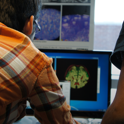
| Synergy of image analysis for animal and human neuroimaging supports translational research on drug abuse G. Gerig, I. Oguz, S. Gouttard, J. Lee, H. An, W. Lin, M. McMurray, K. Grewen, J. Johns, M.A. Styner. In Frontiers in Child and Neurodevelopmental Psychiatry, Vol. 2, Edited by Linda Mayes, pp. 9 pages. 2011. ISSN: 1664-0640 DOI: 10.3389/fpsyt.2011.00053 |
| Efficient Probabilistic and Geometric Anatomical Mapping Using Particle Mesh Approximation on GPUs L.K. Ha, M.W. Prastawa, G. Gerig, J.H. Gilmore, C.T. Silva. In International Journal of Biomedical Imaging, Special Issue in Parallel Computation in Medical Imaging Applications, Vol. 2011, Note: Article ID 572187, pp. 16 pages. 2011. DOI: 10.1155/2011/572187 |
|
Multi-scale Unbiased Diffeomorphic Atlas Construction on Multi-GPUs L.K. Ha, J. Krüger, S. Joshi, C.T. Silva. Vol. 1, Ch. 10, Morgan Kaufmann, pp. 42. 2011. In this chapter, we present a high performance multi-scale 3D image processing framework to exploit the parallel processing power of multiple graphic processing units (Multi-GPUs) for medical image analysis. We developed GPU algorithms and data structures that can be applied to a wide range of 3D image processing applications and efficiently exploit the computational power and massive bandwidth offered by modern GPUs. Our framework helps scientists solve computationally intensive problems which previously required super computing power. To demonstrate the effectiveness of our framework and to compare to existing techniques, we focus our discussions on atlas construction - the application of understanding the development of the brain and the progression of brain diseases. |
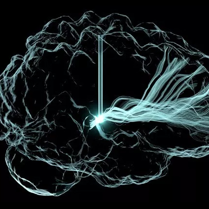
| Adaptive Riemannian Metrics for Improved Geodesic Tracking of White Matter X. Hao, R.T. Whitaker, P.T. Fletcher. In Information Processing in Medical Imaging (IPMI), Lecture Notes in Computer Science (LNCS), Vol. 6801/2011, pp. 13--24. 2011. DOI: 10.1007/978-3-642-22092-0_2 |
| Estimation of Smooth Growth Trajectories with Controlled Acceleration from Time Series Shape Data J. Fishbaugh, S. Durrleman, G. Gerig. In Lecture Notes in Computer Science, LNCS 6892, Springer, pp. 401--408. 2011. DOI: 10.1007/978-3-642-23629-7_49 Longitudinal shape analysis often relies on the estimation of a realistic continuous growth scenario from data sparsely distributed in time. In this paper, we propose a new type of growth model parameterized by acceleration, whereas standard methods typically control the velocity. This mimics the behavior of biological tissue as a mechanical system driven by external forces. The growth trajectories are estimated as smooth flows of deformations, which are twice differentiable. This differs from piecewise geodesic regression, for which the velocity may be discontinuous. We evaluate our approach on a set of anatomical structures of the same subject, scanned 16 times between 4 and 8 years of age. We show our acceleration based method estimates smooth growth, demonstrating improved regularity compared to piecewise geodesic regression. Leave-several-out experiments show that our method is robust to missing observations, as well as being less sensitive to noise, and is therefore more likely to capture the underlying biological growth. Keywords: na-mic |
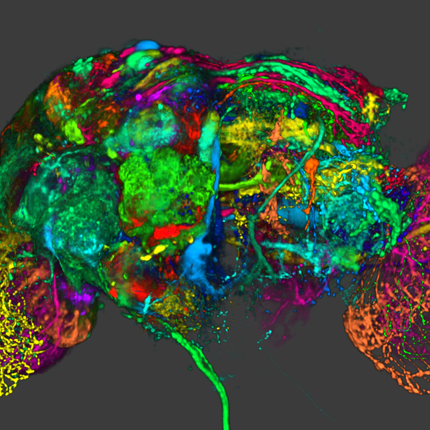
| Three-dimensional reconstruction of serial mouse brain sections using high-resolution large-scale mosaics M.L. Berlanga, S. Phan, E.A. Bushong, S. Lamont, S. Wu, O. Kwon, B.S. Phung, M. Terada, T. Tasdizen, E. Martone, M.H. Ellisman. In Frontiers in Neuroscience Methods, Vol. 5, pp. (published online). March, 2011. DOI: 10.3389/fnana.2011.00017 |
| Detection of Neuron Membranes in Electron Microscopy Images using Multi-scale Context and Radon-like Features M. Seyedhosseini, R. Kumar, E. Jurrus, R. Guily, M. Ellisman, H. Pfister, T. Tasdizen. In Medical Image Computing and Computer-Assisted Intervention – MICCAI 2011, Lecture Notes in Computer Science (LNCS), Vol. 6891, pp. 670--677. 2011. DOI: 10.1007/978-3-642-23623-5_84 |
| A rapid 2-D centerline extraction method based on tensor voting Z. Leng, J.R. Korenberg, B. Roysam, T. Tasdizen. In 2011 IEEE International Symposium on Biomedical Imaging: From Nano to Macro, pp. 1000--1003. 2011. DOI: 10.1109/ISBI.2011.5872570 |
| Quantifying variability in radiation dose due to respiratory-induced tumor motion S.E. Geneser, J.D. Hinkle, R.M. Kirby, Bo Wang, B. Salter, S. Joshi. In Medical Image Analysis, Vol. 15, No. 4, pp. 640--649. 2011. DOI: 10.1016/j.media.2010.07.003 |
| Fast AdaBoost training using weighted novelty selection M. Seyedhosseini, A.R.C. Paiva, T. Tasdizen. In Proc. IEEE Intl. Joint Conf. on Neural Networks, San Jose, CA, USA pp. 1245--1250. August, 2011. In this paper, a new AdaBoost learning framework, called WNS-AdaBoost, is proposed for training discriminative models. The proposed approach significantly speeds up the learning process of adaptive boosting (AdaBoost) by reducing the number of data points. For this purpose, we introduce the weighted novelty selection (WNS) sampling strategy and combine it with AdaBoost to obtain an efficient and fast learning algorithm. WNS selects a representative subset of data thereby reducing the number of data points onto which AdaBoost is applied. In addition, WNS associates a weight with each selected data point such that the weighted subset approximates the distribution of all the training data. This ensures that AdaBoost can trained efficiently and with minimal loss of accuracy. The performance of WNS-AdaBoost is first demonstrated in a classification task. Then, WNS is employed in a probabilistic boosting-tree (PBT) structure for image segmentation. Results in these two applications show that the training time using WNS-AdaBoost is greatly reduced at the cost of only a few percent in accuracy. |
| Point Set Registration Using Havrda–Charvat–Tsallis Entropy Measures N.J. Tustison, S.P. Awate, G. Song, T.S. Cook, J.C. Gee. In IEEE Transactions on Medical Imaging, Vol. 30, No. 2, pp. 451--460. 2011. |
| FADTTS: Functional Analysis of Diffusion Tensor Tract Statistics H. Zhu, L. Kong, R. Li, M.S. Styner, G. Gerig, W. Lin, J.H. Gilmore. In NeuroImage, Vol. 56, No. 3, pp. 1412--1425. 2011. DOI: 10.1016/j.neuroimage.2011.01.075 PubMed ID: 21335092 |
| DTI registration in atlas based fiber analysis of infantile Krabbe disease Y. Wang, A. Gupta, Z. Liu, H. Zhang, M.L. Escolar, J.H. Gilmore, S. Gouttard, P. Fillard, E. Maltbie, G. Gerig, M. Styner. In Neuroimage, pp. (in print). 2011. PubMed ID: 21256236 |
| Twin-Singleton Differences in Neonatal Brain Structure R.C. Knickmeyer, C. Kang, S. Woolson, K.J. Smith, R.M. Hamer, W. Lin, G. Gerig, M. Styner, J.H. Gilmore. In Twin Research and Human Genetics, Vol. 14, No. 3, pp. 268--276. 2011. ISSN: 1832-4274 DOI: 10.1375/twin.14.3.268 |
| Early Brain Overgrowth in Autism Associated with an Increase in Cortical Surface Area Before Age 2 H.C. Hazlett, M. Poe, G. Gerig, M. Styner, C. Chappell, R.G. Smith, C. Vachet, J. Piven. In Arch of Gen Psych, Vol. 68, No. 5, pp. 467--476. 2011. DOI: 10.1001/archgenpsychiatry.2011.39 |
| Optimal data-driven sparse parameterization of diffeomorphisms for population analysis S. Durrleman, M.W. Prastawa, G. Gerig, S. Joshi. In Proceedings of the IPMI 2011 conference, Springer LNCS, Vol. 6801/2011, pp. 123--134. July, 2011. DOI: 10.1007/978-3-642-22092-0_11 PubMed ID: 20516153 |
| Spatial Intensity Prior Correction for Tissue Segmentation in the Developing human Brain S.H. Kim, V. Fonov, J. Piven, J. Gilmore, C. Vachet, G. Gerig, D.L. Collins, M. Styner. In Proceedings of IEEE ISBI 2011, pp. 2049--2052. 2011. DOI: 10.1109/ISBI.2011.5872815 |
| CENTS: Cortical Enhanced Neonatal Tissue Segmentation F. Shi, D. Shen, P.-T. Yap, Y. Fan, J.-Z. Cheng, H. An, L.L. Wald, G. Gerig, J.H. Gilmore, W. Lin. In Human Brain Mapping HBM, Vol. 32, No. 3, Note: ePub 5 Aug 2010, pp. 382--396. March, 2011. DOI: 10.1002/hbm.21023 PubMed ID: 20690143 |
| Multi-scale Series Contextual Model for Image Parsing SCI Technical Report, M. Seyedhosseini, A.R.C. Paiva, T. Tasdizen. No. UUSCI-2011-004, SCI Institute, University of Utah, 2011. |