
Image Analysis
SCI's imaging work addresses fundamental questions in 2D and 3D image processing, including filtering, segmentation, surface reconstruction, and shape analysis. In low-level image processing, this effort has produce new nonparametric methods for modeling image statistics, which have resulted in better algorithms for denoising and reconstruction. Work with particle systems has led to new methods for visualizing and analyzing 3D surfaces. Our work in image processing also includes applications of advanced computing to 3D images, which has resulted in new parallel algorithms and real-time implementations on graphics processing units (GPUs). Application areas include medical image analysis, biological image processing, defense, environmental monitoring, and oil and gas.
Ross Whitaker
Segmentation


Chris Johnson
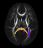
Diffusion Tensor Analysis
Funded Research Projects:
Publications in Image Analysis:

|
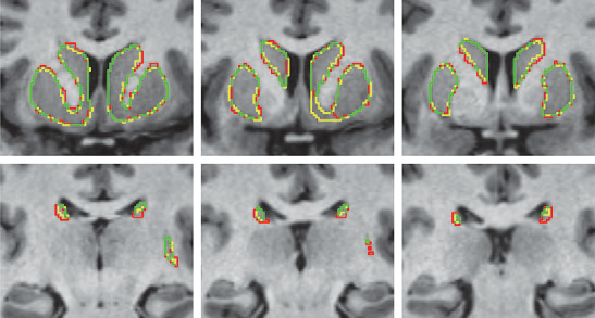
  The DTI Challenge: Toward Standardized Evaluation of Diffusion Tensor Imaging Tractography for Neurosurgery S. Pujol, W. Wells, C. Pierpaoli, C. Brun, J. Gee, G. Cheng, B. Vemuri, O. Commowick, S. Prima, A. Stamm, M. Goubran, A. Khan, T. Peters, P. Neher, K. H. Maier-Hein, Y. Shi, A. Tristan-Vega, G. Veni, R. Whitaker, M. Styner, C.F. Westin, S. Gouttard, I. Norton, L. Chauvin, H. Mamata, G. Gerig, A. Nabavi, A. Golby,, R. Kikinis. In Journal of Neuroimaging, Wiley, August, 2015. DOI: 10.1111/jon.12283 BACKGROUND AND PURPOSE |
Performance of an Efficient Image-registration Algorithm in Processing MR Renography Data, C.C. Conlin, J.L. Zhang, F. Rousset, C. Vachet, Y. Zhao, K.A. Morton, K. Carlston, G. Gerig, V.S. Lee. In J Magnetic Resonance Imaging, July, 2015. DOI: 10.1002/jmri.25000 PURPOSE: |
|
A Kalman Filtering Perspective for Multiatlas Segmentation Y. Gao, L. Zhu, J. Cates, R. S. MacLeod, S. Bouix,, A. Tannenbaum. In SIAM J. Imaging Sciences, Vol. 8, No. 2, pp. 1007-1029. 2015. DOI: 10.1137/130933423 In multiatlas segmentation, one typically registers several atlases to the novel image, and their respective segmented label images are transformed and fused to form the final segmentation. In this work, we provide a new dynamical system perspective for multiatlas segmentation, inspired by the following fact: The transformation that aligns the current atlas to the novel image can be not only computed by direct registration but also inferred from the transformation that aligns the previous atlas to the image together with the transformation between the two atlases. This process is similar to the global positioning system on a vehicle, which gets position by inquiring from the satellite and by employing the previous location and velocity—neither answer in isolation being perfect. To solve this problem, a dynamical system scheme is crucial to combine the two pieces of information; for example, a Kalman filtering scheme is used. Accordingly, in this work, a Kalman multiatlas segmentation is proposed to stabilize the global/affine registration step. The contributions of this work are twofold. First, it provides a new dynamical systematic perspective for standard independent multiatlas registrations, and it is solved by Kalman filtering. Second, with very little extra computation, it can be combined with most existing multiatlas segmentation schemes for better registration/segmentation accuracy. |
|
Modeling Brain Growth and Development N. Sadeghi, J. H. Gilmore , G. Gerig. In Brain, Vol. 1, pp. 429-436. 2015. DOI: 10.1016/B978-0-12-397025-1.00314-6 Early brain development is characterized by rapid organization and structuring. Magnetic resonance–diffusion tensor imaging (MR-DTI) provides the possibility of capturing these changes noninvasively by following individuals longitudinally to better understand departures from normal brain development in subjects at risk for mental illness. This article illustrates the modeling of neurodevelopmental trajectories using a recently developed framework. Descriptions include the estimation of normative models for healthy singletons and twins and a statistical framework to predict development at 2 years of age only based on neonatal image data – a capability with excellent potential for preclinical diagnosis and eventual early therapeutic intervention. |

|
Altered corpus callosum morphology associated with autism over the first 2 years of life J. J. Wolff, G. Gerig, J. D. Lewis, T. Soda, M. A. Styner, C. Vachet, K. N. Botteron, J. T. Elison, S. R. Dager, A. M. Estes, H. C. Hazlett, R. T. Schultz, L. Zwaigenbaum, J. Piven. In Brain, 2015. DOI: 10.1093/brain/awv118 Numerous brain imaging studies indicate that the corpus callosum is smaller in older children and adults with autism spectrum disorder. However, there are no published studies examining the morphological development of this connective pathway in infants at-risk for the disorder. Magnetic resonance imaging data were collected from 270 infants at high familial risk for autism spectrum disorder and 108 low-risk controls at 6, 12 and 24 months of age, with 83% of infants contributing two or more data points. Fifty-seven children met criteria for ASD based on clinical-best estimate diagnosis at age 2 years. Corpora callosa were measured for area, length and thickness by automated segmentation. We found significantly increased corpus callosum area and thickness in children with autism spectrum disorder starting at 6 months of age. These differences were particularly robust in the anterior corpus callosum at the 6 and 12 month time points. Regression analysis indicated that radial diffusivity in this region, measured by diffusion tensor imaging, inversely predicted thickness. Measures of area and thickness in the first year of life were correlated with repetitive behaviours at age 2 years. In contrast to work from older children and adults, our findings suggest that the corpus callosum may be larger in infants who go on to develop autism spectrum disorder. This result was apparent with or without adjustment for total brain volume. Although we did not see a significant interaction between group and age, cross-sectional data indicated that area and thickness differences diminish by age 2 years. Regression data incorporating diffusion tensor imaging suggest that microstructural properties of callosal white matter, which includes myelination and axon composition, may explain group differences in morphology. |
| Investigating maternal brain structure and its relationship to substance use and motivational systems H. J.V. Rutherford, G. Gerig, S. Gouttard, M. N. Potenza, L. C. Mayes. In Yale Journal of Biology and Medicine, in print, 2015. Substance use during pregnancy and the postpartum period may have significant implications for both mother and the developing child. However, the neurobiological basis of the impact of substance use on parenting is less well understood. Here we examined the impact of maternal substance use on cortical gray matter (GM) and white matter volumes, and whether this was associated with individual differences in motivational systems of behavioral activation and inhibition. Mothers were included in the substance-using group if any addictive substance was used during pregnancy and/or in the immediate postpartum period (within 3 months of delivery). GM volume was reduced in substance-using mothers compared to non-substance-using mothers, particularly in frontal brain regions. In substance-using mothers, we also found that frontal GM was negatively correlated with levels of behavioral activation (i.e., the motivation to approach rewarding stimuli). This effect was absent in non-substance-using mothers. Taken together, these findings indicate a reduction in GM volume is associated with substance use, and that frontal GM volumetric differences may be related to approach motivation in substance-using mothers. |

|
Bayesian Principal Geodesic Analysis for Estimating Intrinsic Diffeomorphic Image Variability M. Zhang, P. T. Fletcher. In Medical Image Analysis (accepted), 2015. In this paper, we present a generative Bayesian approach for estimating the low-dimensional latent space of diffeomorphic shape variability in a population of images. We develop a latent variable model for principal geodesic analysis (PGA) that provides a probabilistic framework for factor analysis in the space of diffeomorphisms. A sparsity prior in the model results in automatic selection of the number of relevant dimensions by driving unnecessary principal geodesics to zero. To infer model parameters, including the image atlas, principal geodesic deformations, and the effective dimensionality, we introduce an expectation maximization (EM) algorithm. We evaluate our proposed model on 2D synthetic data and the 3D OASIS brain database of magnetic resonance images, and show that the automatically selected latent dimensions from our model are able to reconstruct unobserved testing images with lower error than both linear principal component analysis (LPCA) in the image space and tangent space principal component analysis (TPCA) in the diffeomorphism space. |
|
Prenatal Drug Exposure Affects Neonatal Brain Functional Connectivity A. P. Salzwedel, K. M. Grewen, C. Vachet, G. Gerig, W. Lin,, W. Gao. In The Journal of Neuroscience, Vol. 35, No. 14, pp. 5860-5869. April, 2015. DOI: 10.1523/JNEUROSCI.4333-14.2015 Prenatal drug exposure, particularly prenatal cocaine exposure (PCE), incurs great public and scientific interest because of its associated neurodevelopmental consequences. However, the neural underpinnings of PCE remain essentially uncharted, and existing studies in school-aged children and adolescents are confounded greatly by postnatal environmental factors. In this study, leveraging a large neonate sample (N = 152) and non-invasive resting-state functional magnetic resonance imaging, we compared human infants with PCE comorbid with other drugs (such as nicotine, alcohol, marijuana, and antidepressant) with infants with similar non-cocaine poly drug exposure and drug-free controls. We aimed to characterize the neural correlates of PCE based on functional connectivity measurements of the amygdala and insula at the earliest stage of development. Our results revealed common drug exposure-related connectivity disruptions within the amygdala–frontal, insula–frontal, and insula–sensorimotor circuits. Moreover, a cocaine-specific effect was detected within a subregion of the amygdala–frontal network. This pathway is thought to play an important role in arousal regulation, which has been shown to be irregular in PCE infants and adolescents. These novel results provide the earliest human-based functional delineations of the neural-developmental consequences of prenatal drug exposure and thus open a new window for the advancement of effective strategies aimed at early risk identification and intervention. |
|
Finite-Dimensional Lie Algebras for Fast Diffeomorphic Image Registration M. Zhang, P. T. Fletcher. In Information Processing in Medical Imaging (IPMI), 2015. This paper presents a fast geodesic shooting algorithm for diffeomorphic image registration. We first introduce a novel finite-dimensional Lie algebra structure on the space of bandlimited velocity fields. We then show that this space can effectively represent initial velocities for diffeomorphic image registration at much lower dimensions than typically used, with little to no loss in registration accuracy. We then leverage the fact that the geodesic evolution equations, as well as the adjoint Jacobi field equations needed for gradient descent methods, can be computed entirely in this finite-dimensional Lie algebra. The result is a geodesic shooting method for large deformation metric mapping (LDDMM) that is dramatically faster and less memory intensive than state-of-the-art methods. We demonstrate the effectiveness of our model to register 3D brain images and compare its registration accuracy, runtime, and memory consumption with leading LDDMM methods. We also show how our algorithm breaks through the prohibitive time and memory requirements of diffeomorphic atlas building. |
Spatio-temporal Image Analysis for Longitudinal and Time-Series Image Data, S. Durrleman, T.P. Fletcher, G. Gerig, M. Niethammer, X. Pennec (Eds.). In Proceedings of the Third International Workshop, STIA 2014, Image Processing, Computer Vision, Pattern Recognition, and Graphics, Vol. 8682, Springer LNCS, 2015. ISBN: 978-3-319-14905-9 This book constitutes the thoroughly refereed post-conference proceedings of the Third |
| Cell tracking using particle filters with implicit convex shape model in 4D confocal microscopy images N. Ramesh, T. Tasdizen. In 2014 IEEE International Conference on Image Processing (ICIP), IEEE, Oct, 2014. DOI: 10.1109/icip.2014.7025089 Bayesian frameworks are commonly used in tracking algorithms. An important example is the particle filter, where a stochastic motion model describes the evolution of the state, and the observation model relates the noisy measurements to the state. Particle filters have been used to track the lineage of cells. Propagating the shape model of the cell through the particle filter is beneficial for tracking. We approximate arbitrary shapes of cells with a novel implicit convex function. The importance sampling step of the particle filter is defined using the cost associated with fitting our implicit convex shape model to the observations. Our technique is capable of tracking the lineage of cells for nonmitotic stages. We validate our algorithm by tracking the lineage of retinal and lens cells in zebrafish embryos. |
Multiatlas Segmentation as Nonparametric Regression S.P. Awate, R.T. Whitaker. In IEEE Trans Med Imaging, April, 2014. PubMed ID: 24802528 This paper proposes a novel theoretical framework to model and analyze the statistical characteristics of a wide range of segmentation methods that incorporate a database of label maps or atlases; such methods are termed as label fusion or multiatlas segmentation. We model these multiatlas segmentation problems as nonparametric regression problems in the high-dimensional space of image patches. We analyze the nonparametric estimator's convergence behavior that characterizes expected segmentation error as a function of the size of the multiatlas database. We show that this error has an analytic form involving several parameters that are fundamental to the specific segmentation problem (determined by the chosen anatomical structure, imaging modality, registration algorithm, and labelfusion algorithm). We describe how to estimate these parameters and show that several human anatomical structures exhibit the trends modeled analytically. We use these parameter estimates to optimize the regression estimator. We show that the expected error for large database sizes is well predicted by models learned on small databases. Thus, a few expert segmentations can help predict the database sizes required to keep the expected error below a specified tolerance level. Such cost-benefit analysis is crucial for deploying clinical multiatlas segmentation systems. |
|
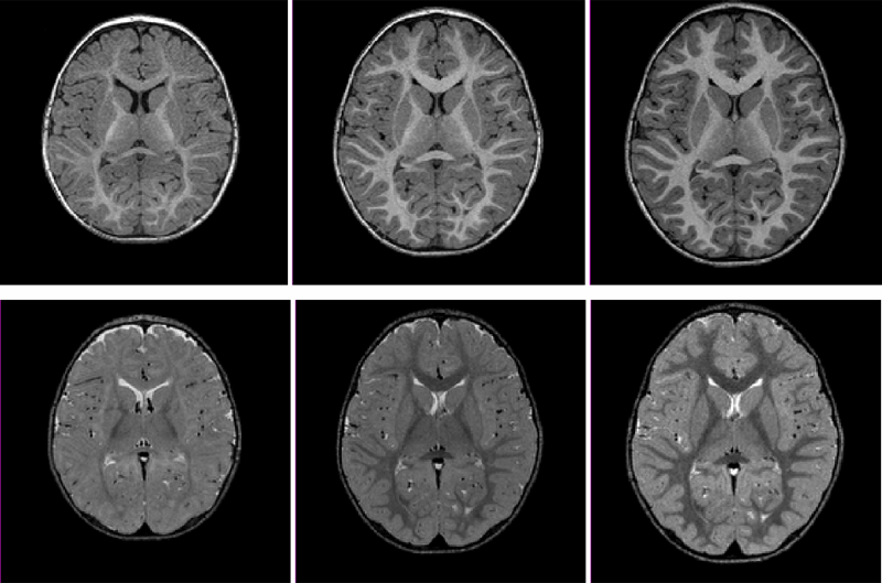
Joint Longitudinal Modeling of Brain Appearance in Multimodal MRI for the Characterization of Early Brain Developmental Processes A. Vardhan, N. Sadeghi, C. Vachet, J. Piven, G. Gerig. In Spatiotemporal Image Analysis for Longitudinal and Time-Series Image Data (STIA'14) , LNCS. MICCAI'14, Springer Verlag, June, 2014. Early brain maturational processes such as myelination manifest as changes in the relative appearance of white-gray matter tissue classes in MR images. Imaging modalities such as T1W (T1-Weighted) and T2W (T2-Weighted) MRI each display specific patterns of appearance change associated with distinct neurobiological components of these maturational processes. In this paper we present a framework to jointly model multimodal appearance changes across time for a longitudinal imaging dataset, resulting in quantitative assessment of the patterns of early brain maturation not yet available to clinicians. We measure appearance by quantifying contrast between white and gray matter in terms of the distance between their intensity distributions, a method demonstrated to be relatively stable to interscan variability. A multivariate nonlinear mixed effects (NLME) model is used for joint statistical modeling of this contrast measure across multiple imaging modalities. The multivariate NLME procedure considers correlations between modalities in addition to intra-modal variability. The parameters of the logistic growth function used in NLME modeling provide useful quantitative information about the timing and progression of contrast change in multimodal datasets. Inverted patterns of relative white-gray matter intensity gradient that are observable in T1W scans with respect to T2W scans are characterized by the SIR (Signal Intensity Ratio). The CONTDIR (Contrast Direction) which measures the direction of the gradient at each time point relative to that in the adult-like scan adds a directional attribute to contrast. The major contribution of this paper is a framework for joint multimodal temporal modeling of white-gray matter MRI contrast change and estimation of subject-specific and population growth trajectories. Results confirm qualitative descriptions of growth patterns in pediatric radiology studies and our new quantitative modeling scheme has the potential to advance understanding of variability of brain tissue maturation and to eventually differentiate normal from abnormal growth for early diagnosis of pathology. |

|
Diffeomorphic Shape Trajectories for Improved Longitudinal Segmentation and Statistics P. Muralidharan, J. Fishbaugh, H.J. Johnson, S. Durrleman, J.S. Paulsen, G. Gerig, P.T. Fletcher. In Proceedings of Medical Image Computing and Computer Assisted Intervention (MICCAI), 2014. Longitudinal imaging studies involve tracking changes in individuals by repeated image acquisition over time. The goal of these studies is to quantify biological shape variability within and across individuals, and also to distinguish between normal and disease populations. However, data variability is influenced by outside sources such as image acquisition, image calibration, human expert judgment, and limited robustness of segmentation and registration algorithms. In this paper, we propose a two-stage method for the statistical analysis of longitu- dinal shape. In the first stage, we estimate diffeomorphic shape trajectories for each individual that minimize inconsistencies in segmented shapes across time. This is followed by a longitudinal mixed-effects statistical model in the second stage for testing differences in shape trajectories between groups. We apply our method to a longitudinal database from PREDICT-HD and demonstrate our ap- proach reduces unwanted variability for both shape and derived measures, such as volume. This leads to greater statistical power to distinguish differences in shape trajectory between healthy subjects and subjects with a genetic biomarker for Huntington's disease (HD). |
|
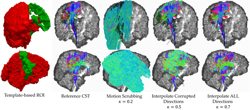
Motion is inevitable: The impact of motion correction schemes on hardi reconstructions S. Elhabian, Y. Gur, J. Piven, M. Styner, I. Leppert, G. Bruce Pike, G. Gerig. In Proceedings of the MICCAI 2014 Workshop on Computational Diffusion MRI, September, 2014. Diffusion weighted imaging (DWI) is known to be prone to artifacts related to motion originating from subject movement, cardiac pulsation and breathing, but also to mechanical issues such as table vibrations. Given the necessity for rigorous quality control and motion correction, users are often left to use simple heuristics to select correction schemes, but do not fully understand the consequences of such choices on the final analysis, moreover being at risk to introduce confounding factors in population studies. This paper reports work in progress towards a comprehensive evaluation framework of HARDI motion correction to support selection of optimal methods to correct for even subtle motion. We make use of human brain HARDI data from a well controlled motion experiment to simulate various degrees of motion corruption. Choices for correction include exclusion or registration of motion corrupted directions, with different choices of interpolation. The comparative evaluation is based on studying effects of motion correction on three different metrics commonly used when using DWI data, including similarity of fiber orientation distribution functions (fODFs), global brain connectivity via Graph Diffusion Distance (GDD), and reproducibility of prominent and anatomically defined fiber tracts. Effects of various settings are systematically explored and illustrated, leading to the somewhat surprising conclusion that a best choice is the alignment and interpolation of all DWI directions, not only directions considered as corrupted. |
|
Prenatal cocaine effects on brain structure in early infancy K. Grewen, M. Burchinal, C. Vachet, S. Gouttard, J.H. Gilmore, W. Lin, J. Johns, M. Elam, G. Gerig. In NeuroImage, Vol. 101, pp. 114--123. November, 2014. DOI: 10.1016/j.neuroimage.2014.06.070 Prenatal cocaine exposure (PCE) is related to subtle deficits in cognitive and behavioral function in infancy, childhood and adolescence. Very little is known about the effects of in utero PCE on early brain development that may contribute to these impairments. The purpose of this study was to examine brain structural differences in infants with and without PCE. We conducted MRI scans of newborns (mean age = 5 weeks) to determine cocaine's impact on early brain structural development. Subjects were three groups of infants: 33 with PCE co-morbid with other drugs, 46 drug-free controls and 40 with prenatal exposure to other drugs (nicotine, alcohol, marijuana, opiates, SSRIs) but without cocaine. Infants with PCE exhibited lesser total gray matter (GM) volume and greater total cerebral spinal fluid (CSF) volume compared with controls and infants with non-cocaine drug exposure. Analysis of regional volumes revealed that whole brain GM differences were driven primarily by lesser GM in prefrontal and frontal brain regions in infants with PCE, while more posterior regions (parietal, occipital) did not differ across groups. Greater CSF volumes in PCE infants were present in prefrontal, frontal and parietal but not occipital regions. Greatest differences (GM reduction, CSF enlargement) in PCE infants were observed in dorsal prefrontal cortex. Results suggest that PCE is associated with structural deficits in neonatal cortical gray matter, specifically in prefrontal and frontal regions involved in executive function and inhibitory control. Longitudinal study is required to determine whether these early differences persist and contribute to deficits in cognitive functions and enhanced risk for drug abuse seen at school age and in later life. |
|
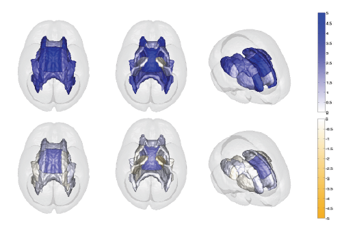
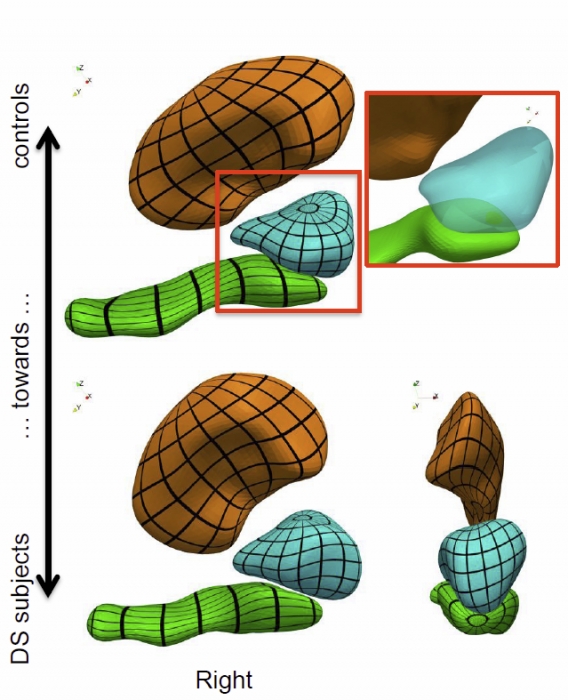
Morphometry of anatomical shape complexes with dense deformations and sparse parameters S. Durrleman, M. Prastawa, N. Charon, J.R. Korenberg, S. Joshi, G. Gerig, A. Trouvé. In NeuroImage, 2014. DOI: 10.1016/j.neuroimage.2014.06.043 We propose a generic method for the statistical analysis of collections of anatomical shape complexes, namely sets of surfaces that were previously segmented and labeled in a group of subjects. The method estimates an anatomical model, the template complex, that is representative of the population under study. Its shape reflects anatomical invariants within the dataset. In addition, the method automatically places control points near the most variable parts of the template complex. Vectors attached to these points are parameters of deformations of the ambient 3D space. These deformations warp the template to each subject’s complex in a way that preserves the organization of the anatomical structures. Multivariate statistical analysis is applied to these deformation parameters to test for group differences. Results of the statistical analysis are then expressed in terms of deformation patterns of the template complex, and can be visualized and interpreted. The user needs only to specify the topology of the template complex and the number of control points. The method then automatically estimates the shape of the template complex, the optimal position of control points and deformation parameters. The proposed approach is completely generic with respect to any type of application and well adapted to efficient use in clinical studies, in that it does not require point correspondence across surfaces and is robust to mesh imperfections such as holes, spikes, inconsistent orientation or irregular meshing. The approach is illustrated with a neuroimaging study of Down syndrome (DS). Results demonstrate that the complex of deep brain structures shows a statistically significant shape difference between control and DS subjects. The deformation-based modelingis able to classify subjects with very high specificity and sensitivity, thus showing important generalization capability even given a low sample size. We show that results remain significant even if the number of control points, and hence the dimension of variables in the statistical model, are drastically reduced. The analysis may even suggest that parsimonious models have an increased statistical performance. The method has been implemented in the software Deformetrica, which is publicly available at www.deformetrica.org.
Keywords: morphometry, deformation, varifold, anatomy, shape, statistics |
| Assessment of white matter microstructure in stroke patients using NODDI G. Adluru, Y. Gur, J. Anderson, L. Richards, N. Adluru, E. DiBella. In Proceedings of the 2014 IEEE Int. Conf. Engineering and Biology Society (EMBC), 2014. Diffusion weighted imaging (DWI) is widely used to study changes in white matter following stroke. In various studies employing diffusion tensor imaging (DTI) and high angular resolution diffusion imaging (HARDI) modalities, it has been shown that fractional anisotropy (FA), mean diffusivity (MD), and generalized FA (GFA) can be used as measures of white matter tract integrity in stroke patients. However, these measures may be non-specific, as they do not directly delineate changes in tissue microstructure. Multi-compartment models overcome this limitation by modeling DWI data using a set of indices that are directly related to white matter microstructure. One of these models which is gaining popularity, is neurite orientation dispersion and density imaging (NODDI). This model uses conventional single or multi-shell HARDI data to describe fiber orientation dispersion as well as densities of different tissue types in the imaging voxel. In this paper, we apply for the first time the NODDI model to 4-shell HARDI stroke data. By computing NODDI indices over the entire brain in two stroke patients, and comparing tissue regions in ipsilesional and contralesional hemispheres, we demonstrate that NODDI modeling provides specific information on tissue microstructural changes. We also introduce an information theoretic analysis framework to investigate the non-local effects of stroke in the white matter. Our initial results suggest that the NODDI indices might be more specific markers of white matter reorganization following stroke than other measures previously used in studies of stroke recovery. |
|
Subject-specific prediction using nonlinear population modeling: Application to early brain maturation from DTI N. Sadeghi, P.T. Fletcher, M. Prastawa, J.H. Gilmore, G. Gerig. In Proceedings of Medical Image Computing and Computer-Assisted Intervention (MICCAI 2014), 2014. The term prediction implies expected outcome in the future, often based on a model and statistical inference. Longitudinal imaging studies offer the possibility to model temporal change trajectories of anatomy across populations of subjects. In the spirit of subject-specific analysis, such normative models can then be used to compare data from new subjects to the norm and to study progression of disease or to predict outcome. This paper follows a statistical inference approach and presents a framework for prediction of future observations based on past measurements and population statistics. We describe prediction in the context of nonlinear mixed effects modeling (NLME) where the full reference population's statistics (estimated fixed effects, variance-covariance of random effects, variance of noise) is used along with the individual's available observations to predict its trajectory. The proposed methodology is generic in regard to application domains. Here, we demonstrate analysis of early infant brain maturation from longitudinal DTI with up to three time points. Growth as observed in DTI-derived scalar invariants is modeled with a parametric function, its parameters being input to NLME population modeling. Trajectories of new subject's data are estimated when using no observation, only the rst or the first two time points. Leave-one-out experiments result in statistics on differences between actual and predicted observations. We also simulate a clinical scenario of prediction on multiple categories, where trajectories predicted from multiple models are classified based on maximum likelihood criteria. |

|
GuideME: Slice-guided Semiautomatic Multivariate Exploration of Volumes L. Zhou, C.D. Hansen. In Computer Graphics Forum, Vol. 33, No. 3, Wiley-Blackwell, pp. 151--160. jun, 2014. DOI: 10.1111/cgf.12371 Multivariate volume visualization is important for many applications including petroleum exploration and medicine. State-of-the-art tools allow users to interactively explore volumes with multiple linked parameter-space views. However, interactions in the parameter space using trial-and-error may be unintuitive and time consuming. Furthermore, switching between different views may be distracting. In this paper, we propose GuideME: a novel slice-guided semiautomatic multivariate volume exploration approach. Specifically, the approach comprises four stages: attribute inspection, guided uncertainty-aware lasso creation, automated feature extraction and optional spatial fine tuning and visualization. Throughout the exploration process, the user does not need to interact with the parameter views at all and examples of complex real-world data demonstrate the usefulness, efficiency and ease-of-use of our method. |