
Image Analysis
SCI's imaging work addresses fundamental questions in 2D and 3D image processing, including filtering, segmentation, surface reconstruction, and shape analysis. In low-level image processing, this effort has produce new nonparametric methods for modeling image statistics, which have resulted in better algorithms for denoising and reconstruction. Work with particle systems has led to new methods for visualizing and analyzing 3D surfaces. Our work in image processing also includes applications of advanced computing to 3D images, which has resulted in new parallel algorithms and real-time implementations on graphics processing units (GPUs). Application areas include medical image analysis, biological image processing, defense, environmental monitoring, and oil and gas.
Ross Whitaker
Segmentation


Chris Johnson
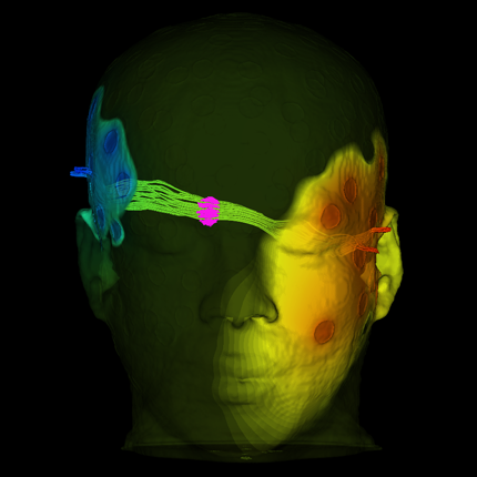
Diffusion Tensor Analysis
Funded Research Projects:
Publications in Image Analysis:
  Learning to Estimate the Composition of a Mixture with Synthetic Data C. Ly, C. Nizinski, C. Vachet, L. McDonald, T. Tasdizen. In Microscopy and Microanalysis, 2021. Identifying the precise composition of a mixed material is important in various applications. For instance, in nuclear forensics analysis, knowing the process history of unknown or illicitly trafficked nuclear materials when they are discovered is desirable to prevent future losses or theft of material from the processing facilities. Motivated by this open problem, we describe a novel machine learning approach to determine the composition of a mixture from SEM images. In machine learning, the training data distribution should reflect the distribution of the data the model is expected to make predictions for, which can pose a hurdle. However, a key advantage of our proposed framework is that it requires reference images of pure material samples only. Removing the need for reference samples of various mixed material compositions reduces the time and monetary cost associated with reference sample preparation and imaging. Moreover, our proposed framework can determine the composition of a mixture composed of chemically similar materials, whereas other elemental analysis tools such as powder X-ray diffraction (p-XRD) have trouble doing so. For example, p-XRD is unable to discern mixtures composed of triuranium octoxide (U3O8) synthesized from different synthetic routes such as uranyl peroxide (UO4) and ammonium diuranate (ADU) [1]. In contrast, our proposed framework can easily determine the composition of uranium oxides mixture synthesized from different synthetic routes, as we illustrate in the experiments. |
| Leveraging Unsupervised Image Registration for Discovery of Landmark Shape Descriptor R. Bhalodia, S. Elhabian, L. Kavan, R. Whitaker. In Medical Image Analysis, Elsevier, pp. 102157. 2021. In current biological and medical research, statistical shape modeling (SSM) provides an essential framework for the characterization of anatomy/morphology. Such analysis is often driven by the identification of a relatively small number of geometrically consistent features found across the samples of a population. These features can subsequently provide information about the population shape variation. Dense correspondence models can provide ease of computation and yield an interpretable low-dimensional shape descriptor when followed by dimensionality reduction. However, automatic methods for obtaining such correspondences usually require image segmentation followed by significant preprocessing, which is taxing in terms of both computation as well as human resources. In many cases, the segmentation and subsequent processing require manual guidance and anatomy specific domain expertise. This paper proposes a self-supervised deep learning approach for discovering landmarks from images that can directly be used as a shape descriptor for subsequent analysis. We use landmark-driven image registration as the primary task to force the neural network to discover landmarks that register the images well. We also propose a regularization term that allows for robust optimization of the neural network and ensures that the landmarks uniformly span the image domain. The proposed method circumvents segmentation and preprocessing and directly produces a usable shape descriptor using just 2D or 3D images. In addition, we also propose two variants on the training loss function that allows for prior shape information to be integrated into the model. We apply this framework on several 2D and 3D datasets to obtain their shape descriptors. We analyze these shape descriptors in their efficacy of capturing shape information by performing different shape-driven applications depending on the data ranging from shape clustering to severity prediction to outcome diagnosis. |
| Impact of scene-specific enhancement spectra on matched filter greenhouse gas retrievals from imaging spectroscopy M. D. Foote, P. E. Dennison, P. R. Sullivan, K. B. O'Neill, A. K. Thorpe, D. R. Thompson, D. H. Cusworth, R. Duren, S. Joshi. In Remote Sensing of Environment, Vol. 264, Elsevier, pp. 112574. 2021. Matched filter techniques have been widely used for retrieval of greenhouse gas enhancements from imaging spectroscopy datasets. While multiple algorithmic techniques and refinements have been proposed, the greenhouse gas target spectrum used for concentration enhancement estimation has remained largely unaltered since the introduction of quantitative matched filter retrievals. The magnitude of retrieved methane and carbon dioxide enhancements, and thereby integrated mass enhancements (IME) and estimated flux of point-source emitters, is heavily dependent on this target spectrum. Current standard use of molecular absorption coefficients to create unit enhancement target spectra does not account for absorption by background concentrations of greenhouse gases, solar and sensor geometry, or atmospheric water vapor absorption. We introduce geometric and atmospheric parameters into the generation of scene-specific unit enhancement spectra to provide target spectra that are compatible with all greenhouse gas retrieval matched filter techniques. Specifically, we use radiative transfer modeling to model four parameters that are expected to change between scenes: solar zenith angle, column water vapor, ground elevation, and sensor altitude. These parameter values are well defined, with low variation within a single scene. A benchmark dataset consisting of ten AVIRIS-NG airborne imaging spectrometer scenes was used to compare IME retrieved using a matched filter algorithm. For methane plumes, IME resulting from use of standard, generic enhancement spectra varied from −22 to +28.7% compared to scene-specific enhancement spectra. Due to differences in spectral shape between the generic and scene-specific enhancement spectra, differences in methane plume IME were linked to surface spectral characteristics in addition to geometric and atmospheric parameters. IME differences were much larger for carbon dioxide plumes, with generic enhancement spectra producing integrated mass enhancements −76.1 to −48.1% compared to scene-specific enhancement spectra. Fluxes calculated from these integrated enhancements would vary by the same percentages, assuming equivalent wind conditions. Methane and carbon dioxide IME were most sensitive to changes in solar zenith angle and ground elevation. We introduce an interpolation approach that can efficiently generate scene-specific unit enhancement spectra for given sets of parameters. Scene-specific target spectra can improve confidence in greenhouse gas retrievals and flux estimates across collections of scenes with diverse geometric and atmospheric conditions. |
| Bridge Simulation and Metric Estimation on Lie Groups Subtitled “arXiv preprint arXiv:2106.03431,” M. H. Jensen, S. Joshi, S. Sommer. 2021. We present a simulation scheme for simulating Brownian bridges on complete and connected Lie groups. We show how this simulation scheme leads to absolute continuity of the Brownian bridge measure with respect to the guided process measure. This result generalizes the Euclidean result of Delyon and Hu to Lie groups. We present numerical results of the guided process in the Lie group $\SO(3)$. In particular, we apply importance sampling to estimate the metric on $\SO(3)$ using an iterative maximum likelihood method. |
| Interactive Analysis for Large Volume Data from Fluorescence Microscopy at Cellular Precision Y. Wan, H.A. Holman, C. Hansen. In Computers & Graphics, Vol. 98, Pergamon, pp. 138-149. 2021. DOI: https://doi.org/10.1016/j.cag.2021.05.006 The main objective for understanding fluorescence microscopy data is to investigate and evaluate the fluorescent signal intensity distributions as well as their spatial relationships across multiple channels. The quantitative analysis of 3D fluorescence microscopy data needs interactive tools for researchers to select and focus on relevant biological structures. We developed an interactive tool based on volume visualization techniques and GPU computing for streamlining rapid data analysis. Our main contribution is the implementation of common data quantification functions on streamed volumes, providing interactive analyses on large data without lengthy preprocessing. Data segmentation and quantification are coupled with brushing and executed at an interactive speed. A large volume is partitioned into data bricks, and only user-selected structures are analyzed to constrain the computational load. We designed a framework to assemble a sequence of GPU programs to handle brick borders and stitch analysis results. Our tool was developed in collaboration with domain experts and has been used to identify cell types. We demonstrate a workflow to analyze cells in vestibular epithelia of transgenic mice. |
| Loon: Using Exemplars to Visualize Large Scale Microscopy Data D. Lange, E. Polanco, R. Judson-Torres, T. Zangle, A. Lex. In OSF Preprints, 2021. Which drug is most promising for a cancer patient? This is a question a new microscopy-based approach for measuring the mass of individual cancer cells treated with different drugs promises to answer in only a few hours. However, the analysis pipeline for extracting data from these images is still far from complete automation: human intervention is necessary for quality control for preprocessing steps such as segmentation, to adjust filters, and remove noise, and for the analysis of the result. To address this workflow, we developed Loon, a visualization tool for analyzing drug screening data based on quantitative phase microscopy imaging. Loon visualizes both, derived data such as growth rates, and imaging data. Since the images are collected automatically at a large scale, manual inspection of images and segmentations is infeasible. However, reviewing representative samples of cells is essential, both for quality control and for data analysis. We introduce a new approach of choosing and visualizing representative exemplar cells that retain a close connection to the low-level data. By tightly integrating the derived data visualization capabilities with the novel exemplar visualization and providing selection and filtering capabilities, Loon is well suited for making decisions about which drugs are suitable for a specific patient. |
| Small-molecule mimicry hunting strategy in the imperial cone snail, Conus imperialis J. P. Torres, Z. Lin, M. Watkins, P. F. Salcedo, R. P. Baskin, S. Elhabian, H. Safavi-Hemami, D. Taylor, J. Tun, G. P. Concepcion, N. Saguil, A. A. Yanagihara, Y. Fang, J. R. McArthur, H. Tae, R. K. Finol-Urdaneta, B. D. Özpolat, B. M. Olivera, E. W. Schmidt. In Science Advances, Vol. 7, No. 11, American Association for the Advancement of Science, 2021. Venomous animals hunt using bioactive peptides, but relatively little is known about venom small molecules and the resulting complex hunting behaviors. Here, we explored the specialized metabolites from the venom of the worm-hunting cone snail, Conus imperialis. Using the model polychaete worm Platynereis dumerilii, we demonstrate that C. imperialis venom contains small molecules that mimic natural polychaete mating pheromones, evoking the mating phenotype in worms. The specialized metabolites from different cone snails are species-specific and structurally diverse, suggesting that the cones may adopt many different prey-hunting strategies enabled by small molecules. Predators sometimes attract prey using the prey’s own pheromones, in a strategy known as aggressive mimicry. Instead, C. imperialis uses metabolically stable mimics of those pheromones, indicating that, in biological mimicry, even the molecules themselves may be disguised, providing a twist on fake news in chemical ecology. |
| Learning Deep Features for Shape Correspondence with Domain Invariance Subtitled “arXiv preprint arXiv:2102.10493,” P. Agrawal, R. T. Whitaker, S. Y. Elhabian. 2021. Correspondence-based shape models are key to various medical imaging applications that rely on a statistical analysis of anatomies. Such shape models are expected to represent consistent anatomical features across the population for population-specific shape statistics. Early approaches for correspondence placement rely on nearest neighbor search for simpler anatomies. Coordinate transformations for shape correspondence hold promise to address the increasing anatomical complexities. Nonetheless, due to the inherent shape-level geometric complexity and population-level shape variation, the coordinate-wise correspondence often does not translate to the anatomical correspondence. An alternative, group-wise approach for correspondence placement explicitly models the trade-off between geometric description and the population's statistical compactness. However, these models achieve limited success in resolving nonlinear shape correspondence. Recent works have addressed this limitation by adopting an application-specific notion of correspondence through lifting positional data to a higher dimensional feature space. However, they heavily rely on manual expertise to create domain-specific features and consistent landmarks. This paper proposes an automated feature learning approach, using deep convolutional neural networks to extract correspondence-friendly features from shape ensembles. Further, an unsupervised domain adaptation scheme is introduced to augment the pretrained geometric features with new anatomies. Results on anatomical datasets of human scapula, femur, and pelvis bones demonstrate that … |
| Image-Based Multiresolution Topology Optimization Using Deep Disjunctive Normal Shape Model V. Keshavarzzadeh, M. Alirezaei, T. Tasdizen, R. M. Kirby. In Computer-Aided Design, Vol. 130, Elsevier, pp. 102947. 2021. We present a machine learning framework for predicting the optimized structural topology design susing multiresolution data. Our approach primarily uses optimized designs from inexpensive coarse mesh finite element simulations for model training and generates high resolution images associated with simulation parameters that are not previously used. Our cost-efficient approach enables the designers to effectively search through possible candidate designs in situations where the design requirements rapidly change. The underlying neural network framework is based on a deep disjunctive normal shape model (DDNSM) which learns the mapping between the simulation parameters and segments of multi resolution images. Using this image-based analysis we provide a practical algorithm which enhances the predictability of the learning machine by determining a limited number of important parametric samples(i.e.samples of the simulation parameters)on which the high resolution training data is generated. We demonstrate our approach on benchmark compliance minimization problems including the 3D topology optimization where we show that the high-fidelity designs from the learning machine are close to optimal designs and can be used as effective initial guesses for the large-scale optimization problem. |
Detection and segmentation in microscopy images, N. Ramesh, T. Tasdizen. In Computer Vision for Microscopy Image Analysis, Academic Press, pp. 43-71. 2021. DOI: 10.1016/B978-0-12-814972-0.00003-5 The plethora of heterogeneous data generated using modern microscopy imaging techniques eliminates the possibility of manual image analysis for biologists. Consequently, reliable and robust computerized techniques are critical to analyze microscopy data. Detection problems in microscopy images focuses on accurately identifying the objects of interest in an image that can be used to investigate hypotheses about developmental or pathological processes and can be indicative of prognosis in patients. Detection is also considered to be the preliminary step for solving subsequent problems, such as segmentation and tracking for various biological applications. Segmentation of the desired structures and regions in microscopy images require pixel-level labels to uniquely identify the individual structures and regions with contours for morphological and physiological analysis. Distributions of features extracted from the segmented regions can be used to compare normal versus disease or normal versus wild-type populations. Segmentation can be considered as a precursor for solving classification, reconstruction, and tracking problems in microscopy images. In this chapter, we discuss how the field of microscopic image analysis has progressed over the years, starting with traditional approaches and then followed by the study of learning algorithms. Because there is a lot of variability in microscopy data, it is essential to study learning algorithms that can adapt to these changes. We focus on deep learning approaches with convolutional neural networks (CNNs), as well as hierarchical methods for segmentation and detection in optical and electron microscopy images. Limitation of training data is one of the significant problems; hence, we explore solutions to learn better models with minimal user annotations. |
| Leveraging 31 Million Google Street View Images to Characterize Built Environments and Examine County Health Outcomes Q. C Nguyen, J. M. Keralis, P. Dwivedi, A. E. Ng, M. Javanmardi, S. Khanna, Y. Huang, K. D. Brunisholz, A. Kumar, T. Tasdizen. In Public Health Reports, Vol. 136, No. 2, SAGE Publications, pp. 201-211. 2021. DOI: doi.org/10.1177/0033354920968799 Objectives Methods
We leveraged computer vision and Google Street View images accessed from December 15, 2017, through July 17, 2018, to detect features of the built environment (presence of a crosswalk, non–single-family home, single-lane roads, and visible utility wires) for 2916 US counties. We used multivariate linear regression models to determine associations between features of the built environment and county-level health outcomes (prevalence of adult obesity, prevalence of diabetes, physical inactivity, frequent physical and mental distress, poor or fair self-rated health, and premature death [in years of potential life lost]).Results
Compared with counties with the least number of crosswalks, counties with the most crosswalks were associated with decreases of 1.3%, 2.7%, and 1.3% of adult obesity, physical inactivity, and fair or poor self-rated health, respectively, and 477 fewer years of potential life lost before age 75 (per 100 000 population). The presence of non–single-family homes was associated with lower levels of all health outcomes except for premature death. The presence of single-lane roads was associated with an increase in physical inactivity, frequent physical distress, and fair or poor self-rated health. Visible utility wires were associated with increases in adult obesity, diabetes, physical and mental distress, and fair or poor self-rated health.Conclusions
The use of computer vision and big data image sources makes possible national studies of the built environm
|
| Lessons learned towards the immediate delivery of massive aerial imagery to farmers and crop consultants A. A. Gooch, S. Petruzza, A. Gyulassy, G. Scorzelli, V. Pascucci, L. Rantham, W. Adcock, C. Coopmans. In Autonomous Air and Ground Sensing Systems for Agricultural Optimization and Phenotyping VI, Vol. 11747, International Society for Optics and Photonics, pp. 22 -- 34. 2021. DOI: 10.1117/12.2587694 In this paper, we document lessons learned from using ViSOAR Ag Explorer™ in the fields of Arkansas and Utah in the 2018-2020 growing seasons. Our insights come from creating software with fast reading and writing of 2D aerial image mosaics for platform-agnostic collaborative analytics and visualization. We currently enable stitching in the field on a laptop without the need for an internet connection. The full resolution result is then available for instant streaming visualization and analytics via Python scripting. While our software, ViSOAR Ag Explorer™ removes the time and labor software bottleneck in processing large aerial surveys, enabling a cost-effective process to deliver actionable information to farmers, we learned valuable lessons with regard to the acquisition, storage, viewing, analysis, and planning stages of aerial data surveys. Additionally, with the ultimate goal of stitching thousands of images in minutes on board a UAV at the time of data capture, we performed preliminary tests for on-board, real-time stitching and analysis on USU AggieAir sUAS using lightweight computational resources. This system is able to create a 2D map while flying and allow interactive exploration of the full resolution data as soon as the platform has landed or has access to a network. This capability further speeds up the assessment process on the field and opens opportunities for new real-time photogrammetry applications. Flying and imaging over 1500-2000 acres per week provides up-to-date maps that give crop consultants a much broader scope of the field in general as well as providing a better view into planting and field preparation than could be observed from field level. Ultimately, our software and hardware could provide a much better understanding of weed presence and intensity or lack thereof. |
| Structural Connectome Atlas Construction in the Space of Riemannian Metrics Subtitled “arXiv,” K. M. Campbell, H. Dai, Z. Su, M. Bauer, P. T. Fletcher, S. C. Joshi. 2021. The structural connectome is often represented by fiber bundles generated from various types of tractography. We propose a method of analyzing connectomes by representing them as a Riemannian metric, thereby viewing them as points in an infinite-dimensional manifold. After equipping this space with a natural metric structure, the Ebin metric, weapply object-oriented statistical analysis to define an atlas as the Fŕechet mean of a population of Riemannian metrics. We demonstrate connectome registration and atlas formation using connectomes derived from diffusion tensors estimated from a subset of subjects from the Human Connectome Project. |
| Physics Informed Convex Artificial Neural Networks (PICANNs) for Optimal Transport based Density Estimation Subtitled “arXiv,” A. Singh, M. Bauer, S. Joshi. 2021. Optimal Mass Transport (OMT) is a well studied problem with a variety of applications in a diverse set of fields ranging from Physics to Computer Vision and in particular Statistics and Data Science. Since the original formulation of Monge in 1781 significant theoretical progress been made on the existence, uniqueness and properties of the optimal transport maps. The actual numerical computation of the transport maps, particularly in high dimensions, remains a challenging problem. By Brenier's theorem, the continuous OMT problem can be reduced to that of solving a non-linear PDE of Monge-Ampere type whose solution is a convex function. In this paper, building on recent developments of input convex neural networks and physics informed neural networks for solving PDE's, we propose a Deep Learning approach to solve the continuous OMT problem. |
Determining uranium ore concentrates and their calcination products via image classification of multiple magnifications, C. Ly, C. Vachet, I. Schwerdt, E. Abbott, A. Brenkmann, L.W. McDonald, T. Tasdizen. In Journal of Nuclear Materials, 2020. Many tools, such as mass spectrometry, X-ray diffraction, X-ray fluorescence, ion chromatography, etc., are currently available to scientists investigating interdicted nuclear material. These tools provide an analysis of physical, chemical, or isotopic characteristics of the seized material to identify its origin. In this study, a novel technique that characterizes physical attributes is proposed to provide insight into the processing route of unknown uranium ore concentrates (UOCs) and their calcination products. In particular, this study focuses on the characteristics of the surface structure captured in scanning electron microscopy (SEM) images at different magnification levels. Twelve common commercial processing routes of UOCs and their calcination products are investigated. Multiple-input single-output (MISO) convolution neural networks (CNNs) are implemented to differentiate the processing routes. The proposed technique can determine the processing route of a given sample in under a second running on a graphics processing unit (GPU) with an accuracy of more than 95%. The accuracy and speed of this proposed technique enable nuclear scientists to provide the preliminary identification results of interdicted material in a short time period. Furthermore, this proposed technique uses a predetermined set of magnifications, which in turn eliminates the human bias in selecting the magnification during the image acquisition process. |
Evaluation and validation of off-the-shelf statistical shape modeling tools in clinical applications Anupama Goparaju. School of Computing, University of Utah, 2019. Statistical shape modeling (SSM) enables quantitative analysis of anatomical shapes. SSM is widely used in biology and medicine to model anatomies and their shape variability within populations. The technological advancements of in vivo images have led to the development of various open-source tools that can automate statistical analysis of shapes. These tools are based on different modeling approaches and assumptions to accomplish the same objective. However, little work has been done in the systematic evaluation and validation of SSM tools in clinical applications that rely on morphometric quantifications. |
Adversarial regression training for visualizing the progression of chronic obstructive pulmonary disease with chest x-rays, R.B. Lanfredi, J.D. Schroeder, C. Vachet, T. Tasdizen. In Arxiv, In International Conference on Medical Image Computing and Computer-Assisted Intervention, 2019. Knowledge of what spatial elements of medical images deep learning methods use as evidence is important for model interpretability, trustiness, and validation. There is a lack of such techniques for models in regression tasks. We propose a method, called visualization for regression with a generative adversarial network (VR-GAN), for formulating adversarial training specifically for datasets containing regression target values characterizing disease severity. We use a conditional generative adversarial network where the generator attempts to learn to shift the output of a regressor through creating disease effect maps that are added to the original images. Meanwhile, the regressor is trained to predict the original regression value for the modified images. A model trained with this technique learns to provide visualization for how the image would appear at different stages of the disease. We analyze our method in a dataset of chest x-rays associated with pulmonary function tests, used for diagnosing chronic obstructive pulmonary disease (COPD). For validation, we compute the difference of two registered x-rays of the same patient at different time points and correlate it to the generated disease effect map. The proposed method outperforms a technique based on classification and provides realistic-looking images, making modifications to images following what radiologists usually observe for this disease. Implementation code is available athttps://github.com/ricbl/vrgan. |
| A Cooperative Autoencoder for Population-Based Regularization of CNN Image Registration R. Bhalodia, S. Y. Elhabian, L. Kavan, R. T. Whitaker. In Medical Image Computing and Computer Assisted Intervention – MICCAI 2019, In Medical Image Computing and Computer Assisted Intervention -- MICCAI 2019, Springer International Publishing, pp. 391--400. 2019. Spatial transformations are enablers in a variety of medical image analysis applications that entail aligning images to a common coordinate systems. Population analysis of such transformations is expected to capture the underlying image and shape variations, and hence these transformations are required to produce anatomically feasible correspondences. This is usually enforced through some smoothness-based generic metric or regularization of the deformation field. Alternatively, population-based regularization has been shown to produce anatomically accurate correspondences in cases where anatomically unaware (i.e., data independent) regularization fail. Recently, deep networks have been used to generate spatial transformations in an unsupervised manner, and, once trained, these networks are computationally faster and as accurate as conventional, optimization-based registration methods. However, the deformation fields produced by these networks require smoothness penalties, just as the conventional registration methods, and ignores population-level statistics of the transformations. Here, we propose a novel neural network architecture that simultaneously learns and uses the population-level statistics of the spatial transformations to regularize the neural networks for unsupervised image registration. This regularization is in the form of a bottleneck autoencoder, which learns and adapts to the population of transformations required to align input images by encoding the transformations to a low dimensional manifold. The proposed architecture produces deformation fields that describe the population-level features and associated correspondences in an anatomically relevant manner and are statistically compact relative to the state-of-the-art approaches while maintaining computational efficiency. We demonstrate the efficacy of the proposed architecture on synthetic data sets, as well as 2D and 3D medical data. |
| Which Two-dimensional Radiographic Measurements of Cam Femoroacetabular Impingement Best Describe the Three-dimensional Shape of the Proximal Femur? P. R. Atkins, Y. Shin, P. Agrawal, S. Y. Elhabian, R. T. Whitaker, J. A. Weiss, S. K. Aoki, C. L. Peters, A. E. Anderson. In Clinical Orthopaedics and Related Research, Vol. 477, No. 1, 2019. BACKGROUND: |
Identifying surface morphological characteristics to differentiate between mixtures of U3O8 synthesized from ammonium diuranate and uranyl peroxide, S. T. Heffernan, N. Ly, B. J. Mower, C. Vachet, I. J. Schwerdt, T. Tasdizen, L. W. McDonald IV. In Radiochimica Acta, 2019. In the present study, surface morphological differences of mixtures of triuranium octoxide (U3O8), synthesized from uranyl peroxide (UO4) and ammonium diuranate (ADU), were investigated. The purity of each sample was verified using powder X-ray diffractometry (p-XRD), and scanning electron microscopy (SEM) images were collected to identify unique morphological features. The U3O8 from ADU and UO4 was found to be unique. Qualitatively, both particles have similar features being primarily circular in shape. Using the morphological analysis of materials (MAMA) software, particle shape and size were quantified. UO4 was found to produce U3O8 particles three times the area of those produced from ADU. With the starting morphologies quantified, U3O8 samples from ADU and UO4 were physically mixed in known quantities. SEM images were collected of the mixed samples, and the MAMA software was used to quantify particle attributes. As U3O8 particles from ADU were unique from UO4, the composition of the mixtures could be quantified using SEM imaging coupled with particle analysis. This provides a novel means of quantifying processing histories of mixtures of uranium oxides. Machine learning was also used to help further quantify characteristics in the image database through direct classification and particle segmentation using deep learning techniques based on Convolutional Neural Networks (CNN). It demonstrates that these techniques can distinguish the mixtures with high accuracy as well as showing significant differences in morphology between the mixtures. Results from this study demonstrate the power of quantitative morphological analysis for determining the processing history of nuclear materials. |